What is retinitis pigmentosa?
Retinitis pigmentosa (RP) is the name of a group of inherited, degenerative eye diseases of the retina that results in progressive loss of vision. The retina is the light-sensitive layer at the back of the eye. It is estimated that retinitis pigmentosa affects about 1 in 4,000 people worldwide [1].
Get Involved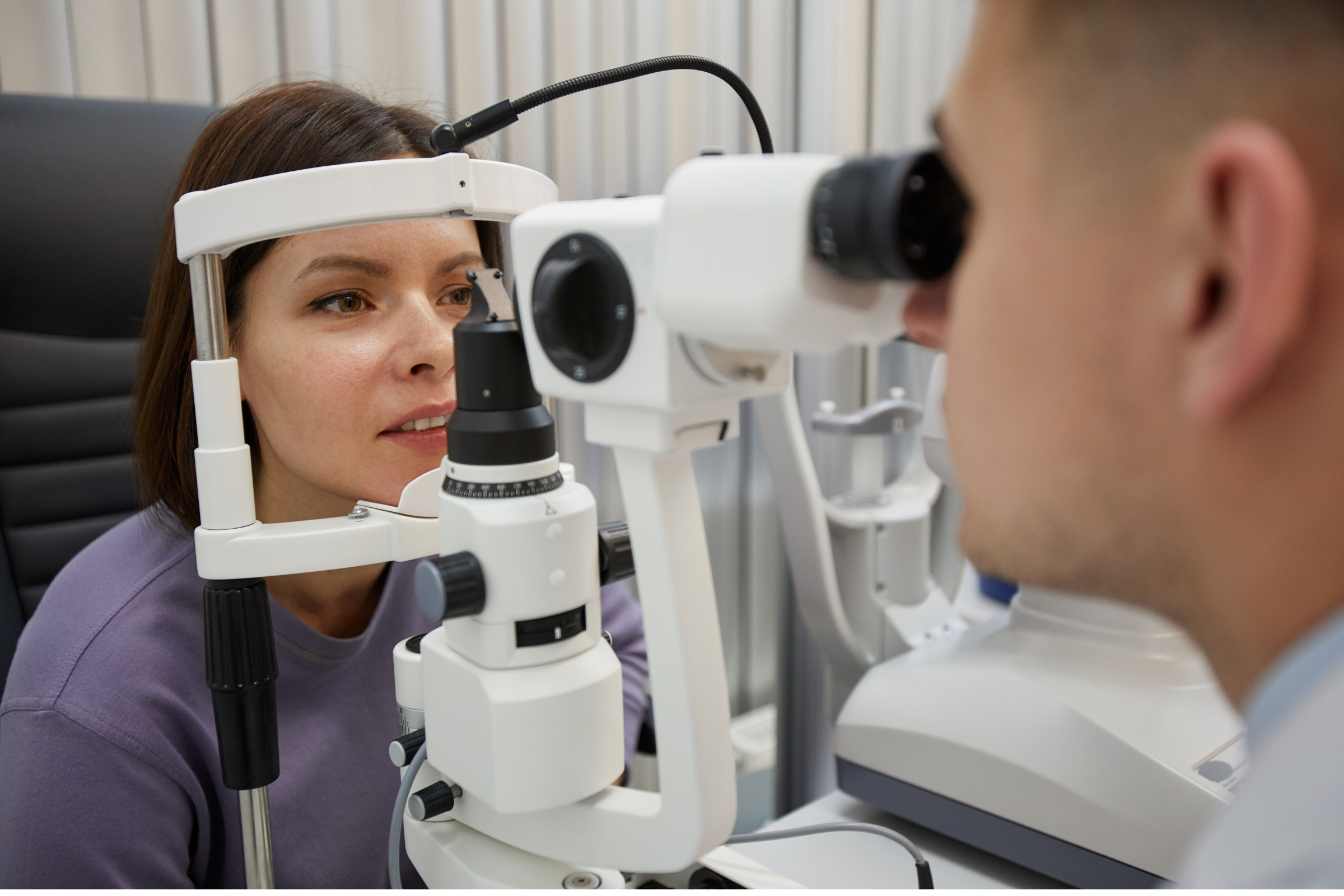
The retina can be compared to a camera, where light-sensitive cells (photoreceptors called rods and cones) send information to the brain for processing.
Rods are scattered throughout the retina, especially around the periphery, and are essential for seeing in dim light and sensing motion.
Cones are found in the central retina in an area called the macula; they are needed for seeing in bright light, colour and sharp detail.
The most common type of RP generally causes night-blindness initially.
This means it is harder to see or takes longer to adjust to dim light, typically more than the usual 15-30 minutes. Some people have trouble with glare from bright lights.
After the onset of night-blindness the person experiences enlarging blind spots in the peripheral vision [2] (ability to see from the corners of the eyes).
Eventually this leads to “tunnel vision”, and in most people, total loss of vision over several decades [3].
RP has many subtypes and symptoms, even varying within families.
Some people with RP have genes that cause problems with other organs, known as syndromes (the most common associated syndrome being Usher syndrome which also causes deafness) [4].
Although retinitis can be caused by faulty genes, about half of people with retinitis pigmentosa have no family history [4].
Diagnosis of RP is made by symptoms, physical examination, and tests.
Cataracts (clouding of lens) are a complication seen in nearly half of people with RP [5], and can require surgery at ages as young as 30-60 years [1].
If macular oedema occurs, central vision can become distorted.
Diagnosis of retinitis pigmentosa [1]
Eye examination
Eye chart to check clarity of vision
Colour vision
Visual field (blind spot) test
Fundus exam – the lens and back of the eye are viewed by the doctor using a slit lamp or ophthalmoscope. Dark spots due to pigment, and other changes may be seen
Dark adaptometry – following exposure to bright light, the patient sits in the dark for 30 minutes. The minimum brightness of light able to be seen is measured intermittently
Fundus autofluorescence (FAF) imaging
Blue light is used to take a detailed picture of the retina
Optical coherence tomography (OCT)
The patient sits in front of OCT machine using a chin rest whilst pictures of the eye are taken for 5-10 minutes
Full-field flash electroretinography (ffERG) [6]
A recording electrode is gently positioned on the surface of the eye or the skin of the lower eyelid.
Measurements are taken of the photoreceptors’ electrical responses during periods of dark then light
Genetic test
Blood or saliva sample
Important for diagnosis and planning management
Although there is no cure for retinitis pigmentosa, treatment options and aids are available.
Treatment options for retinitis pigmentosa [3,4,7,8]
Low vision aids / Rehabilitation training
Magnifying aids for close-up activities
Mobile device apps
Anti-glare sunglasses
Avoidance of unnecessary bright light
Assistive devices such as specialised portable headsets that produce magnified images
Orientation/mobility training
Counselling
Can help to talk through the implications of diagnosis
Nutrition
Depending on type of RP, your specialist may recommend vitamin A or E supplements
Cataract treatment
Cataract removal and artificial lens replacement
Macular oedema treatment
Medication
Experimental therapies
Studies are being conducted into gene therapy, retinal cell transplant and retinal prostheses
1. Hong Y, Li H, Sun Y, Ji Y. A Review of Complicated Cataract in Retinitis Pigmentosa: Pathogenesis and Cataract Surgery. (CC BY 4.0). J Ophthalmol [Internet]. 2020 [cited 2022 May 3];2020:1–14. Available from: https://www.hindawi.com/journals/joph/2020/6699103/#abstract
2. Retinitis pigmentosa. [Internet]. Source: MedlinePlus, National Library of Medicine. 2010 [cited 2022 May 3]. Available from: https://medlineplus.gov/genetics/condition/retinitis-pigmentosa/#references
3. Hamel C. Retinitis pigmentosa. (CC BY 2.0). Orphanet J Rare Dis [Internet]. 2006 Oct 11 [cited 2022 May 2];1(40):1–12. Available from: https://ojrd.biomedcentral.com/articles/10.1186/1750-1172-1-40
4. NORD (National Organization for Rare Disorders). Retinitis Pigmentosa [Internet]. 2021 [cited 2022 May 2]. Available from: https://rarediseases.org/rare-diseases/retinitis-pigmentosa/
5. Fujiwara K, Ikeda Y, Murakami Y, Funatsu J, Nakatake S, Tachibana T, et al. Risk Factors for Posterior Subcapsular Cataract in Retinitis Pigmentosa. Invest Ophthalmol Vis Sci [Internet]. 2017 May 1 [cited 2022 May 3];58(5):2534–7. Available from: https://iovs.arvojournals.org/article.aspx?articleid=2626379
6. Asanad S, Karanjia R. Full-Field Electroretinogram. (CC BY 4.0) [Internet]. StatPearls – NCBI Bookshelf. 2021 [cited 2022 May 3]. Available from: https://www.ncbi.nlm.nih.gov/books/NBK557483/#!po=95.4545
7. Retinitis Pigmentosa [Internet]. Courtesy: National Eye Institute, National Institutes of Health (NEI/NIH). 2022 [cited 2022 May 2]. Available from: https://www.nei.nih.gov/learn-about-eye-health/eye-conditions-and-diseases/retinitis-pigmentosa
8. Low Vision [Internet]. Courtesy: National Eye Institute, National Institutes of Health (NEI/NIH). 2022 [cited 2022 May 3]. Available from: https://www.nei.nih.gov/learn-about-eye-health/eye-conditions-and-diseases/low-vision
Medical Disclaimer
This article contains general information relating to a medical condition. Such information is provided for informational purposes only and does not replace medical advice given by your healthcare professional.
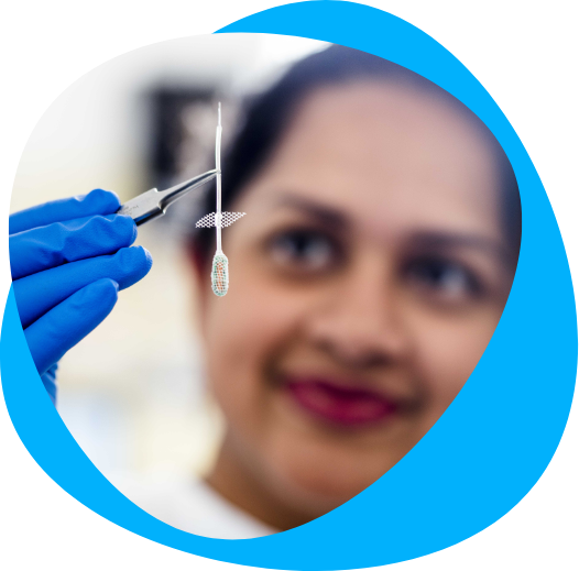
Research at the Bionics Institute into the Bionic Eye
Bionics Institute researchers worked with collaborators on a successful clinical trial of the second-generation Australian bionic eye in 2021.
• The bionic eye is an implant inserted behind the retina that is attached to a video camera built into a pair of glasses.
• The camera converts images into electrical impulses that activate retinal cells using 44 electrodes, designed and manufactured at the Bionics Institute.
• The clinical trial involving four recipients successfully demonstrated that this device is safe and provides significant improvement to quality of life and functional vision.
• In partnership with Bionic Vision Technologies, the next step is to initiate worldwide clinical trials.
Want to support the future of research like this? Early-stage research for life-changing devices is made possible by donations from our supporters. Your support today could turn the seed of an idea into a new treatment in the future. Find out how you can support research innovation here.
Your support today could change the lives of people with vision loss around the world.
Want to support the future of research like this?
Early-stage research for life-changing devices like this is made possible by donations from our supporters.rnYour support today could turn the seed of an idea into a new treatment in the future.rnrnFind out how you can support research innovation here.