Latest News
Heart attack prediction using AI is on the horizon
Ground-breaking AI research was on show at the Bionics Institute 2022 Graeme Clark Oration
Revolutionary technology designed to accurately predict heart attacks was showcased at Melbourne’s premier science event, the Bionics Institute 2022 Graeme Clark Oration in July 2022.
Developed by international researcher Professor Natalia Trayanova (Johns Hopkins University, USA), the artificial intelligence and bioengineering tool could prove to be life-saving for more than four million Australians affected by cardiovascular disease (CVD).
Using data-driven machine learning and biophysics-based modelling, Professor Trayanova has created ‘digital heart twins’ – virtual replicas of a person’s heart that can be used to forecast progress of heart disease, estimate the risk of heart attacks and inform treatment decisions.
“My AI technology uses algorithms created from MRIs and PET scans, in combination with deep learning of clinical data, to predict a patient’s risk of sudden cardiac death over a period of ten years,” Professor Trayanova said.
The application of Professor Trayanova’s technology could be key to reducing the burden on our country’s healthcare system, as CVD costs the Australian economy almost $12 billion annually (2018-2019).
She explained: “I envisage a future where re-hospitalisations and repeat procedures are reduced; shifting heart disease treatment options from being based on the state of the patient today, to optimising the state of the patient for the future.”
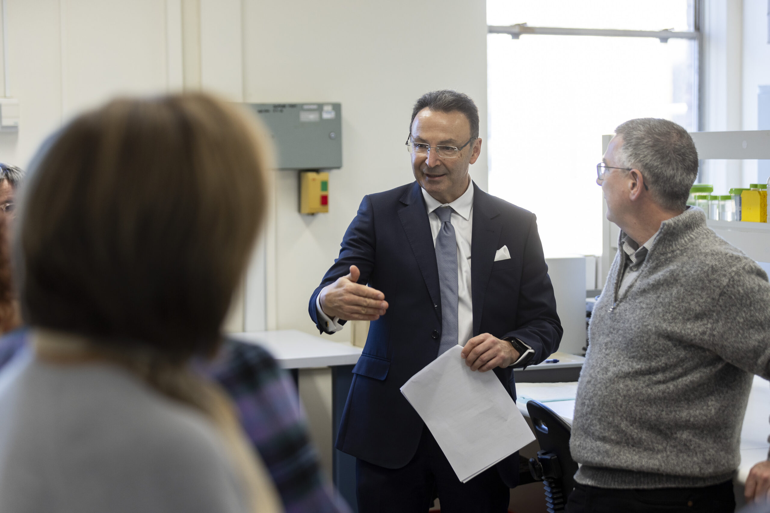
Bionics Institute CEO Mr Robert Klupacs says the Institute was proud to be principal sponsor of the 2022 Graeme Clark Oration, a prestigious science event with a long history of showcasing eminent biomedical scientists.
“Over 1000 Melburnians have registered to join us the Oration and learn about the future of heart healthcare from Professor Trayanova – a pioneer in the field of computational cardiology. This is an event not to be missed,” he said.
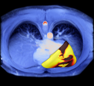
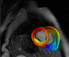
Image, left: A virtual heart model is developed using MRI and PET scans. Bands of colour signify the electrical wave of an irregular heartbeat. Photo courtesy of Professor Natalia Trayanova.
Image, right: A virtual heart is developed using scans of a patient’s heart (blue). Areas where disrupted electrical activity is present (arrhythmia) are represented by yellow and red. Photo courtesy of Professor Natalia Trayanova.


